What evidence did the NIH study provide to show that AI/ML can successfully diagnose Polycystic Ovary Syndrome?

A recent study by the National Institutes of Health found that Artificial intelligence (AI) and machine learning (ML) can be used to detect and diagnose Polycystic Ovary Syndrome (PCOS). PCOS is the most common hormone disorder among women between the ages of 15 and 45. The researchers reviewed published scientific studies that used AI/ML to analyze data to diagnose and classify PCOS. They concluded that AI/ML-based programs were successful in detecting PCOS.

“Given the large burden of under- and misdiagnosed PCOS in the community and its potentially serious outcomes, we wanted to identify the utility of AI/ML in the identification of patients that may be at risk for PCOS,” said Janet Hall, M.D., senior investigator and endocrinologist at the National Institute of Environmental Health Sciences (NIEHS), part of NIH, and a study co-author. “The effectiveness of AI and machine learning in detecting PCOS was even more impressive than we had thought.”

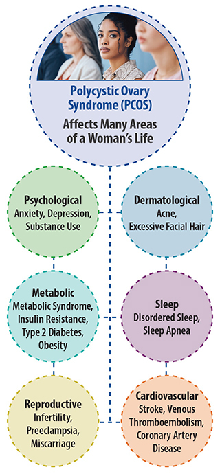
Polycystic ovary syndrome (PCOS) is a hormonal disorder that affects the proper functioning of the ovaries. In many cases, it is accompanied by higher levels of testosterone. This condition can lead to a range of symptoms, including irregular periods, acne, excessive facial hair growth, or baldness. Women with PCOS are more likely to develop type 2 diabetes, as well as sleep, psychological, cardiovascular, and other reproductive disorders such as uterine cancer and infertility.

“PCOS can be challenging to diagnose given its overlap with other conditions,” said Skand Shekhar, M.D., senior author of the study and assistant research physician and endocrinologist at the NIEHS.

“These data reflect the untapped potential of incorporating AI/ML in electronic health records and other clinical settings to improve the diagnosis and care of women with PCOS,” said Shekhar.

The diagnosis of polycystic ovary syndrome (PCOS) is based on standardized criteria that include clinical features, laboratory findings, and radiological evidence. However, PCOS is often difficult to diagnose because some of its symptoms can overlap with other conditions. To improve the accuracy of diagnosis, researchers suggest integrating large population-based studies with electronic health datasets and using machine learning (ML) to identify sensitive diagnostic biomarkers. ML is a type of artificial intelligence (AI) that can process large amounts of data, such as electronic health records. 

In a recent study, researchers conducted a systematic review of peer-reviewed studies published in the last 25 years that used AI/ML to diagnose PCOS. They screened 135 studies and included 31 in their analysis. The studies were all observational and assessed the use of AI/ML technologies for diagnosing PCOS. Ultrasound images were used in about half of the studies, and the average age of the participants was 29. 

The accuracy of PCOS diagnosis using AI/ML varied across the studies. Among the 10 studies that used standardized diagnostic criteria, the accuracy of detection ranged from 80-90%. The findings suggest that AI/ML can be a valuable tool for diagnosing difficult-to-diagnose disorders like PCOS.

“Across a range of diagnostic and classification modalities, there was an extremely high performance of AI/ML in detecting PCOS, which is the most important takeaway of our study,” said Shekhar. The use of AI/ML-based programs has the potential to significantly improve our ability to detect women with PCOS early, which can lead to cost savings and lessen the burden of PCOS on patients and the health system. Further studies with strong validation and testing practices will enable a smooth integration of AI/ML for chronic health conditions. NIEHS clinical studies are currently underway to understand and detect PCOS. To learn more, you can join an NIEHS study.

The findings of the NIH study are clear: AI/ML can be used to successfully diagnose Polycystic Ovary Syndrome. This is a major breakthrough in the medical field, as it demonstrates the potential of AI/ML to improve the accuracy and speed of diagnosis for this and other conditions. With further research and development, AI/ML could revolutionize the way medical professionals diagnose and treat patients.

Grants: This work was supported by the Intramural Research Program of the NIH/National Institute of Environmental Health Sciences (ZIDES102465 and ZIDES103323).

Jamie Spangler  IMAGE CREDIT: WILL KIRK / JOHNS HOPKINS UNIVERSITY
Jamie Spangler IMAGE CREDIT: WILL KIRK / JOHNS HOPKINS UNIVERSITY

Sprangler unlocks the power of nature with 'protein legos'

Breakthrough research has potential implications for the treatment of injuries

Johns Hopkins engineers have helped develop and characterize an artificial protein that triggers the same response in the human body as its natural counterpart, a breakthrough that not only has the potential to facilitate the design of drugs to accelerate healing but also sheds light on the mechanisms behind various diseases.

"It's protein Legos, essentially," said team leader Jamie Spangler, an assistant professor of chemical and biomolecular engineering and biomedical engineering. "We know what the different pieces look like, and we put them together in an arrangement that is predicted to look like the protein we're trying to mimic. As far as the body is concerned, this newly created protein is as genuine as the one that occurs in nature."

The synthetic protein, called Neo-4, mimics the function of the natural protein interleukin-4 (IL-4): a crucial player in immune system regulation. White blood cells release IL-4 in response to a range of immune triggers, from allergic inflammation to muscle injuries. IL-4 can then attach to various receptors on cells throughout the body. However, when IL-4 is directly injected as a drug, it can bind to unintended cells, causing unwanted side effects.

"If you give someone IL-4 it just acts on everything," said Zachary Bernstein, team member and PhD candidate in biomedical engineering. "But that makes it difficult for therapeutic use. Neo-4 is more specific and only activates immunologically relevant cells."

Neo-4 attaches to a narrower range of cells than IL-4, a characteristic that the researchers say could make it a promising candidate for future drug development. For instance, a torn anterior cruciate ligament (ACL) is a common season-ending sports injury. Cytokines like Neo-4 has the potential to speed up the healing of torn ACLs and other damaged ligaments and muscles.

"These are computationally designed proteins that behave like proteins in nature but have better properties," Spangler said. "That means we can build these robust, hyper-stable proteins to do whatever we want. The hope is that we can use this mimetic to deliver IL-4 in a way that is safer and more robust than the natural cytokine, which could help with its therapeutic advancement."

Huilin Yang, a graduate of the doctoral program in chemical and biomolecular engineering contributed to this research.

The use of "protein legos" to enhance the function of natural proteins is a promising development in the field of biochemistry. This technique has the potential to revolutionize the way scientists approach protein engineering, and could lead to the development of new treatments for diseases, as well as new materials and technologies. By continuing to explore the possibilities of this technique, researchers can create a better future for all.

This project was supported by the National Institute of Health and the Emerson Collective Cancer Research Fund, Bruce and Jeannie Nordstrom, and Patty and Jimmy Barrier Gift for the Institute for Protein Design Fund and the National Cancer Institute.

Biomedical engineers and applied mathematicians at Brown created a machine learning algorithm that uses computational topology to study how these cells organize themselves into tissue-like architectures.

Scientists utilize machine learning to uncover the organization of cells and the crucial role topology plays in the process

The research can aid in understanding how cells organize during embryonic development and the consequences of errors in this process.

Embryonic development is a crucial process for the growth and development of living organisms. The process involves the organization of cells in the right way, at the right place and time, to form healthy tissue. Failure in this process can lead to birth defects, impaired tissue regeneration, or cancer. Therefore, understanding how different cell types organize themselves into a complex tissue architecture is a vital question in developmental biology.

For the past few years, a group of Brown University scientists have been using topology, a branch of mathematics, to help the field get closer to understanding an elusive process.

A group of biomedical engineers and applied mathematicians have developed a machine learning algorithm using computational topology to analyze the shapes and spatial patterns of embryos. This algorithm helps study how cells organize themselves into tissue-like structures. In a recent study, the team took this system to the next level by enabling it to study how various types of cells assemble themselves.

“In tissues, there may be differences in how one cell adheres to the same cell type, relative to how it adheres to a different cell type,” said Ian Y. Wong, an associate professor in the School of Engineering who helped develop the algorithm. “There's this interesting question of how these cells know exactly where to end up within a given tissue, which is often spatially compartmentalized into distinct regions.”

During embryonic development in animals, the cells in the outer layer form the skin, the middle layer forms muscle and bone, and the innermost layer forms the liver or lungs. The cells in each layer have an affinity for each other, which means that they preferentially stick together, separating from cells in other layers that go on to form other parts of the body.

In the 1970s, researchers discovered that cells in frog embryos could be gently separated and, when mixed back together, would spontaneously rearrange into their original organization. This occurs because cells have different affinities for each other, and as they cluster and assemble, certain topological patterns of linkages and loops are preserved.

“In the context of these spatial arrangements of tissues, you can learn a lot from what's there, but also from what’s not there at the same time,” said Dhananjay Bhaskar, a recent Brown Ph.D. graduate.

The Brown researchers demonstrated in 2021 how their method can profile the topological traits of a single cell type that organizes into various spatial configurations and make predictions on it.

Originally, the system had a major issue - it was a slow and laborious process. The algorithm compared topological features one by one with other sets of cell positions to determine their differences or similarities. This process took several hours, hindering the algorithm's full potential in understanding how cells assemble themselves. Additionally, it made it difficult to accurately compare what happens when conditions change - a crucial aspect in investigating what goes wrong.

A new study has introduced a method to overcome the limitation of comparing large datasets of cell positions. The research team used persistence images, which are standardized picture-like formats to represent topological features, making it easier to compare these features. They also trained algorithms to generate "digital fingerprints" that capture the key topological features of the data, which allows researchers to classify thousands of simulations of cell organization into similar patterns without human input. This significantly reduces the computation time from hours to seconds. The researchers aim to infer the rules that govern how different cell types arrange themselves based on the final pattern. By tinkering with how certain cells are more or less adhesive, they can identify how and when dramatic alterations occur in tissue architecture.

The process has potential applications in understanding abnormal developmental processes and laboratory experiments involving drug-induced changes to cell migration and adhesion.

“If you can see a certain pattern, we can use our algorithm to tell why that pattern emerges,” Bhaskar said. “In a way, it’s telling us the rules of the game when it comes to cells assembling themselves.”

The research carried out by the scientists has shown the effectiveness of topology in providing a deeper comprehension of how cells organize themselves. This study has the potential to unlock new paths for further investigation into the mechanisms of cellular organization, which could lead to fresh insights into the development and functioning of living organisms. By utilizing topology to investigate the intricate nature of cellular organization, this research has made a valuable contribution to the field of biology and has the potential to transform our understanding of the basic processes of life.